Medical Application of Multi-camera Systems
Bedsore Analysis Using MEDPHOS Photogrammatic System
Medical applications such as bedsore analysis often require 3D surface reconstruction of the human body. Major surface reconstruction techniques are based on mechanical digitizers, laser scanners and vision systems, including photogrammetry. Multi-camera systems can greatly reduce ambiguity resulting from loss of information associated with the perspective mapping of a 3D scene onto a 2D image. The authors present three and four-camera photogrammetric systems for the analysis of bedsores.
A bedsore occurs when sustained pressure of bedclothes against the skin causes local obstruction to the blood supply, resulting in damage and potential loss of the affected tissue. Bedsores affect approximately 20% of hospitalised patients. The cost in both human suffering and financial terms is high; in the US the care of pressure ulcers costs US$ 3 to US$ 5 billion annually, whilst in the UK this figure is currently £300 million, estimated soon to reach £500 million. Both suffering and cost of treatment may be greatly reduced when the ulcer and any changes resulting from therapy can be accurately and reliably measured. Regular objective measurement enables medical staff to accurately assess the progress of wound healing and to improve the treatment strategy. Any measuring technique should preferably be non-invasive so as to avoid damage, infection and pain.
Measurement Techniques
The current clinical method is to place a transparent acetate sheet on the wound and count the number of its bounding squares; this provides a measure of area. Volume is estimated by filling the wound with saline; the volume dispensed from the syringe equals the wound volume. These methods are inaccurate, unreliable and involve contact with the open wound. The non-invasive technique of laser scanning requires a still object during the scan period, which cannot be guaranteed for dynamic living objects like human bodies. Photogrammetry enables recording of objects in a split second and is widely used for non-invasive, quantitative and precise analysis of parts of the human body. For some applications, conventional stereo matching algorithms have their limitations with respect to reliability of match and accuracy. In recent years multi-camera configurations have been proposed to solve some of these limitations for medical applications.
- A multi-camera system has been developed at the Swiss Federal Institute of Technology in Switzerland for 3D face-feature extraction and reconstruction.
- The Aristotle University of Thessaloniki University in Greece has developed a video camera system for the extraction of 3D surface models of the human back.
- The Delft University of Technology in The Netherlands has developed a three-camera system armed with structured light for the measurement of the shape and volume of wounds, to support bedsore analysis.
Three-camera System
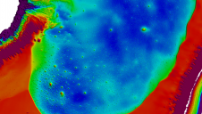
The three-camera photogrammetric system developed in Delft uses a texture projector and is based on least-squares bundle adjustment applied to three stereo pairs provided by three cameras. Figure 1 shows the reconstructed surface model of a large abdominal wound processed with this system. However, intensity-based image matching is not ideal for wound measurement, because:
- human skin is relatively uniform and featureless, so measurement of points on its surface is difficult or impossible
- a wound surface is wet, resulting in specular reflections which disturb the Lambertian condition necessary for intensity-based matching and thus reducing its reliability
- reliable results require an approximation of match positions in the order of a few pixels
- intensity-based methods cannot altogether cope with images of differing radiometric and geometric properties.
Four-camera System
Using four cameras rather than three improves completeness, precision and reliability at little extra cost. We therefore extended the above system to four cameras, calling it MEDPHOS (MEDical PHOtogrammetric System) and basing it on the robust, geometric constraint available in a multiply calibrated camera set-up.
Incorporating the three-focal constraint that takes advantage of epipolar geometry reduces problems caused by intensity-based matching. Moreover, further disambiguation is achieved by projecting a dot pattern over the wound the shape, size and density of which has been optimised. Robust extraction and processing of the dots is done with sophisticated image-processing procedures designed by us. These include homomorphic adjustment for compensation of non-uniform illumination and specular reflectance of the wound surface, watershed segmentation to detect overlapping projected dots and ‘top-hat’ filtering to separate foreground pattern objects from the uneven background. The introduction of a projector calibration reduces remaining ambiguities in match.
Concluding Remarks
MEDPHOS is tailored for bedsore reconstruction, although it may certainly be useful for other applications in medicine and industry. The Faculty of Engineering, University of Tehran provided financial support for construction of the four-camera system.
Further Reading

Value staying current with geomatics?
Stay on the map with our expertly curated newsletters.
We provide educational insights, industry updates, and inspiring stories to help you learn, grow, and reach your full potential in your field. Don't miss out - subscribe today and ensure you're always informed, educated, and inspired.
Choose your newsletter(s)